
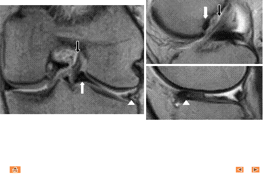
Double ACL sign
Coronal gradient-echo and sagittal T2 MR images
demonstrate displaced lateral meniscal fragment in the intercondylar notch (white
arrow), anterior and lateral to the anterior cruciate ligament (ACL, black arrow). The displaced fragment mimics the ACL. Truncated lateral meniscus is
also demonstrated (white arrowhead).