![]()
![]()
![]()
![]()
![]()
![]()
Atlas of Signs in Musculoskeletal Radiology is approved by the ARRS (American Roentgen Ray Society) and is included in AJR Webreview
A. Gentili,MD, M. Beller, MD, S. Masih, MD, L.L. Seeger, MD
![]()
|
Atlas of Signs in Musculoskeletal Radiology is approved by the ARRS (American Roentgen Ray Society) and is included in AJR WebreviewA. Gentili,MD, M. Beller, MD, S. Masih, MD, L.L. Seeger, MD
|
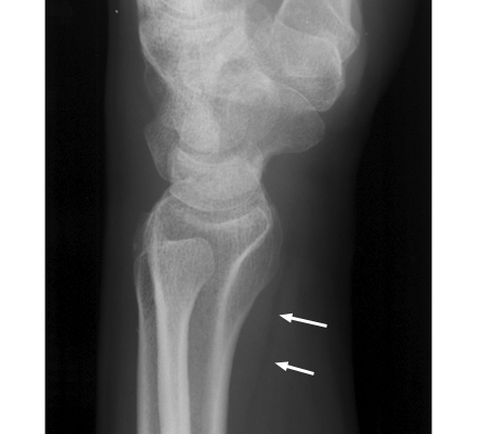
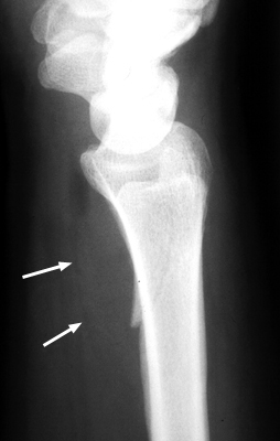
Diagnosis:Wrist Fracture Discussion:A. Normal pronator fat pad. B. Displaced pronator fat pad. Lateral wrist x-ray, a reveals abnormal bulging of a fat plane adjacent to an area of underlying osseous injury. The ventral bulging of the fat plane overlying the pronator quadratus muscle has been coined the "pronator sign." Although it typically means underlying fracture is present, it may also be seen in simple soft-tissue injury of the same region. It is a good sign and should alert the radiologist of possible fracture when present.
References:
|
|
Atlas of Signs in Musculoskeletal Radiology is approved by the ARRS (American Roentgen Ray Society) and is included in AJR WebreviewA. Gentili,MD, M. Beller, MD, S. Masih, MD, L.L. Seeger, MD
|