 CPPD DISEASE
(PSEUDOGOUT) calcium pyrophosphate
dihydrate deposition within cartilage and peri-articular structures.
CPPD DISEASE
(PSEUDOGOUT) calcium pyrophosphate
dihydrate deposition within cartilage and peri-articular structures.
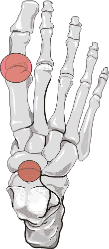
1. Distribution:
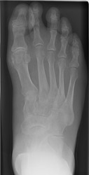
First metatarsophalangeal joint involvement is most common in forefoot. The
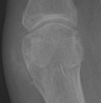
talonavicular joint space is commonly affected with hind foot involvement.
The distribution pattern for CPPD is usually bilateral, and may be symmetric
or asymmetric.
2. Erosion pattern:
Variable, inconsistent osteophyte formation can occur with this disease. Numerous,
large subchondral cysts may progress to micro fractures, collapse, and formation
of intra-articular bodies. Calcific tendonitis (especially of the Achilles
tendon and plantar aponeourosis), or metatarsophalangeal joint capsular or
synovial calcifications may be present. Associated calcifications of the bursa,
ligaments, and fibrocartilaginous structures of the foot are less common.
3. Differential diagnosis:
Joint destruction and cyst formation may resemble osteoarthritic findings.
Talonavicular joint fragmentation may mimic neuropathic arthritis. Synovial
calcifications and soft tissue swelling may be confused with gout. Tendinis
calcifications may mimic calcific tendonitis secondary to calcium hydroxyapatite
crystal deposition disease, and these conditions are commonly co-existent.