![]()
![]()
![]()
![]()
![]()
![]()
![]()
![]()
![]()
![]()
![]()
![]()
![]()
|
|
|
t
t
GoutDeposition of urate and CPPD crystals in periarticular tissues. Usually
present as soft tissue swelling and sharply defined punched out erosions with
overhanging margins.
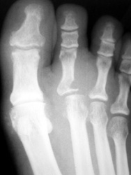
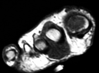
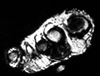
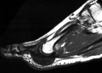
44years old male with rapidly enlarging mass in the foot. Radiographs demonstrates soft tissue mass between the 2nd and 3rd toes,with no calcification.Well defined erosion is noted at the radial aspect of the base of proximal phalanx of 2nd toe. Mild soft tissue swelling with faint calcification is also seen at 1st MTP joint ,with small erosion medial aspect of 1st Mt head. MR demonstrates mass is isointense to hypointense on T1W, and T2W images, with minimum bone marrow edema at the base of proximal phalanx of 2nd toe. Erosion is well seen at the base of proximal phalanx of 2nd toe on T1W images. |
|
|